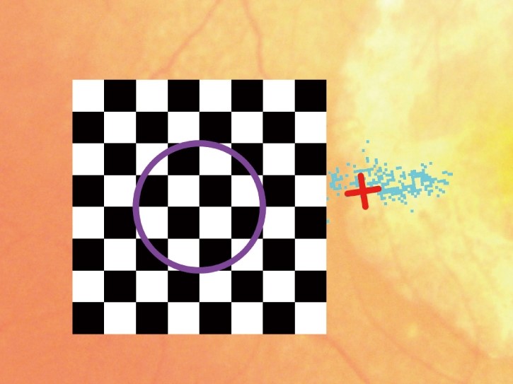
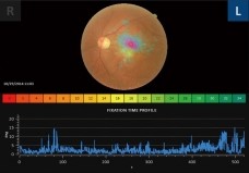
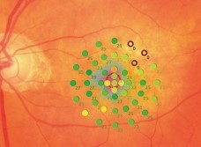
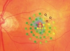
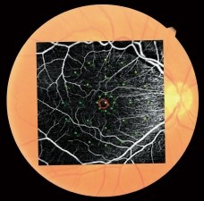
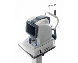
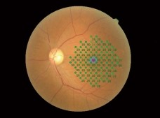
Auto tracking and auto alignment functions provide more accurate measurements increasing patient and operator comfort and efficiency.These functions allow easy follow-up and reduce variations between examiners, resulting in well-aligned follow up exams.Feedback exam for visual rehabilitation The visual rehabilitation mode trains low-vision patients who have lost foveal fixation to relocate their preferred retinal locus (PRL) to a different region, called the trained retinal locus (TRL). The TRL is predetermined by a physician, and fixation rehabilitation allows the patient better functional vision (i.e. reading speed) due to increased fixation stability and visual outcomes.Active flickering pattern stimulation and cheery music create an effective and pleasant training experience for the patient.Fixation testThe MP-3 can measure fixation and determine the preferred retinal locus, simply by having the patient fixate on a target. Any change in fixation can be compared pre- and post-treatment because the patient’s eye is constantly tracked during microperimetry. This test allows evaluation of fixation in patients with central visual field defects and determines whether fixation improved after treatment.
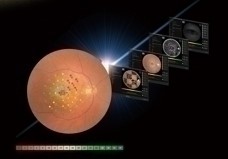
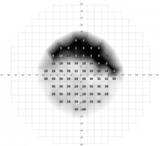
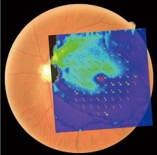
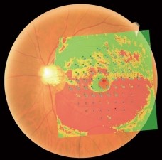
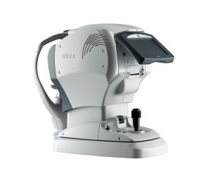
 The MP-3 has a wider range of stimulus intensity, from 0 to 34 dB, compared to the MP-1. The MP-3 measures perimetric threshold values, even for normal eyes. A maximum stimulus luminance of 10,000 asb* allows evaluation of low-sensitivity.*In accordance with ISO12866 measurement methods
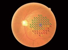