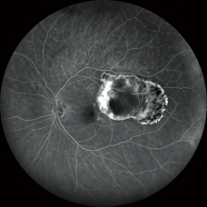
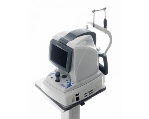
163° ultra wide field imageThe clear image of the entire 163° field of view enables detailed evaluation of pathologies from the fovea to the extreme periphery.(Ultra wide field imaging is available with the optional wide-field adapter.)
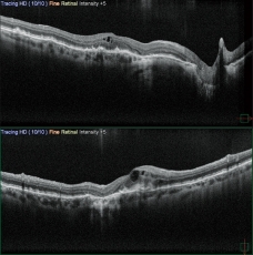
Ultra 4K HD and averaging function for unparalleled clarity
4,096 x 4,096 pixel imaging captures every detail of the retina and choroid. Additionally, zooming in allows high magnification, clear visualization of subtle changes in pathology, and resolution of the fine details of capillaries.New FlexTrack algorithm corrects image distortion due to unstable fixation and enhances averaging quality.
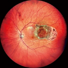
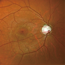
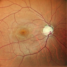
Unsurpassed color
Three separate RGB detectors simultaneously scan different depths of retina with red, green, and blue wavelengths. A color histogram is available for fine adjustment based on pathology or practitioner preference.
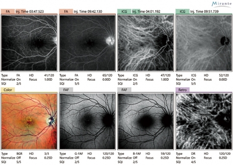
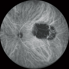
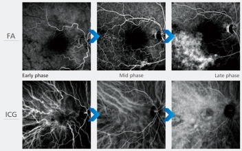
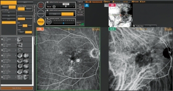
FA and ICG
HD dynamic angiogram
Videos can be recorded at a maximum of 1,024 x 1,024 pixels for up to 120 seconds. Multiple short videos can be recorded during the same measurement.
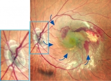
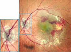
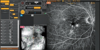
Simultaneous FA and ICG
The Mirante allows simple, simultaneous acquisition of FA and ICG images.
The live IR monitoring enables alignment prior to fluorescence emission and reduces in the risk of missing the very early phase of angiography.
The Auto gain control (AGC) simultaneously adjusts contrast of each FA and ICG image, making the imaging of dynamic blood flow a very simple procedure.
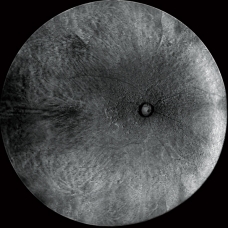
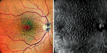
Retro mode
Retro mode is a unique non-invasive technique for detecting pathologic changes in the choroid.
This imaging modality uses scattered IR light to detect abnormal reflection in the choroid caused by drusen, edema and other subtle chorioretinal pathologies