Product Information
Overview
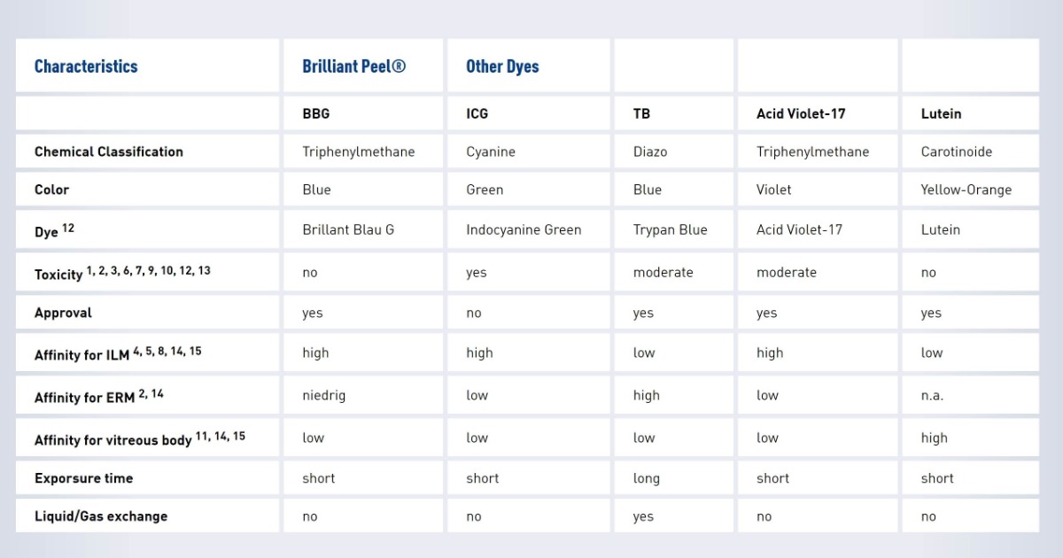
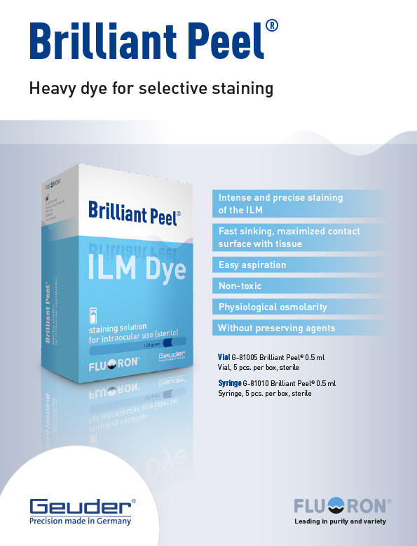
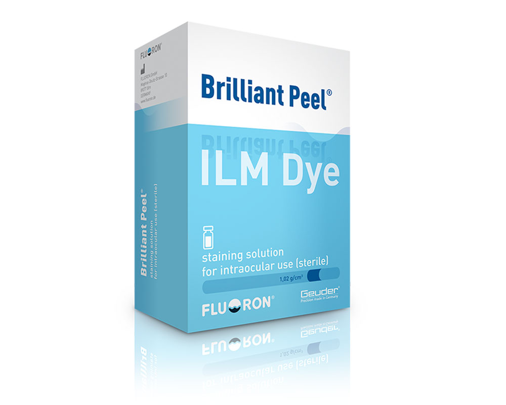
Composition and properties of Brilliant Peel®
Composition of one 0.5 ml syringe / vial:0,125 mg Brilliant Blue G0,95 mg Na2HPO4 x 2 H2O0,15 mg NaH2PO4 x 2 H2O4,1 mg NaCl0,065 ml D2Oad 0,5 ml water for injection purposesConcentration:0,25 g/l Brilliant Blue GDensity:1,02 g/cm3
Use
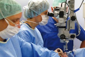
Brilliant Peel® was developed for specific staining of the inner limiting membrane(ILM). Specific staining of the ILM allows it to be clearly differentiated from the underlying retinal tissue and the epiretinal membranes. The surgically demanding removal of the ILM thus becomes easier and safer.
Literature:
1 Lüke C, et al.: Retinal tolerance to dyes, Br J Ophthalmol, 2005, 89, 1188-11912 Haritoglou C, et al.: Färbetechniken in der Makulachirurgie, Ophthalmologe, 2006, 103, 927-9343 Ueno A, et al.: Biocompatibility of Brilliant Blue G in a rat model of subretinal injection, Retina, 2007, 27, 499-5044 Enaida H, et al.: Brilliant Blue G selectively stains the internal limiting membrane – Brilliant Blue G assisted membrane peeling, Retina, 2006, 26, 631 – 6365 Enaida H, et al.: Preclinical investigation of internal limiting membrane staining and peeling using intravitreal Brilliant Blue G, Retina, 2006, 26, 623-6306 Hisatomi T, et al.: Staining ability and biocompatibility of Brilliant Blue G – preclinical study of Brilliant Blue G as an adjunct for capsular staining, Arch Ophthalmol, 2006, 124, 514-5197 Goldman JM, et al.: Adjunct devices for managing challenging cases in cataract surgery – capsular staining and ophthalmic viscosurgical devices, Curr Opin Ophthalmol, 2007, 18, 52-578 Meyer CH, et al.: Historical considerations in applying vital dyes in vitreoretinal surgery: from early experiments to advanced chromovitrectomy, Expert Rev.Ophthalmol., 2007, 71-779 Hiebl W, et al.: Substances for staining biological tissues: use of dyes in ophthalmology, Klin Monatsbl Augenh, 2005, 222, 309-31110 Frank Schuettauf, Christos Haritoglou, Christian A. May, Robert Rejdak, Anna Mankowska, Wolfgang Freyer, Kirsten Eibl, Eberhart Zrenner, Anselm Kampik and Sebastian Thaler, Administration of Novel Dyes for Intraocular Surgery: An In Vivo Toxicity Animal Study, Invest Ophthalmol Vis Sci. 2006; 47:3573–357811 Rodrigues EB, et al.: Vital dyes for chromovitrectomy, Curr Opin Ophthalmol, 2007 May; 18(3):179-8712 Furlani BA, et al.: Lutein and zeaxanthin toxicity with and without brilliant blue in rabbits. J Ocul Pharmacol Ther. 2014 Sep; 30(7):559-66. doi: 10.1089/jop.2013.0171. Epub 2014 Jun 5.13 Tura A, et al.: Testing the effects of the dye acid violet-17 on retinal function for an intraocular application in vitreo-retinal surgery. Graefes Arch Clin Exp Opthalmol. 2014 Dec; 252(12):1927-37. doi: 10.1007/s00417-014-2761-9. Epub 2014 Sep 14.14 Patent DE102012103097 A115 Sousa-Martins D, et al.: Use of lutein and zeaxanthin alone or combined with Brilliant Blue to identify intraocular structures intraoperatively. Retina. 2012 Jul; 32(7):1328-36. doi: 10.1097/IAE.0b013e318239e2b6.